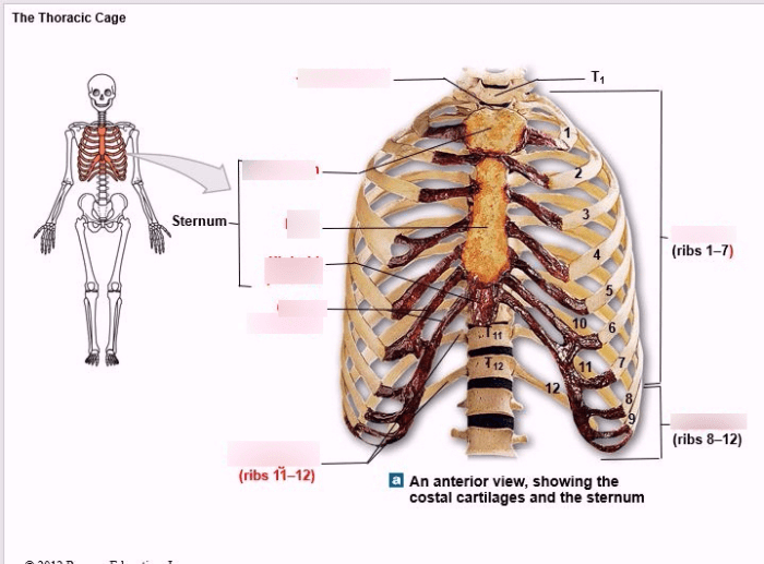
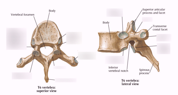
Art-labeling activity the thoracic cage – Embark on an art-labeling expedition that unveils the intricacies of the thoracic cage, a skeletal framework that safeguards vital organs and facilitates respiration. Through this immersive activity, we delve into the anatomical structures, artistic interpretations, and educational value of this remarkable region.
From ancient depictions to modern medical applications, the thoracic cage has captivated artists and scientists alike. Prepare to explore its fascinating history, unravel its anatomical complexities, and discover the transformative power of art in enhancing our understanding of human biology.
Art-Labeling Activity: The Thoracic Cage: Art-labeling Activity The Thoracic Cage

Art-labeling activities are an effective tool for enhancing anatomical knowledge and comprehension. By engaging with visual representations of anatomical structures, students can gain a deeper understanding of their location, shape, and function.
Labeling Techniques, Art-labeling activity the thoracic cage
- Freehand Labeling:Students draw labels directly onto the illustration, providing a personalized and creative approach.
- Sticky Note Labeling:Labels are written on sticky notes and placed on the illustration, allowing for flexibility and easy removal.
- Digital Labeling:Labels are added using digital tools, providing precision and the ability to zoom in for detailed viewing.
Anatomical Structures
| Type | Structure | Location | Shape | Function |
|---|---|---|---|---|
| Bone | Sternum | Anterior midline | Flat, elongated | Protects heart and lungs |
| Bone | Ribs | Attached to sternum and vertebrae | Curved, flattened | Protect thoracic organs |
| Bone | Vertebrae | Posterior | Stacked, cylindrical | Support and protect spinal cord |
| Muscle | Intercostal Muscles | Between ribs | Thin, fan-shaped | Facilitate breathing |
| Nerve | Phrenic Nerve | Innervates diaphragm | Long, slender | Controls breathing |
Artistic Interpretations
Artists throughout history have depicted the thoracic cage in various ways, reflecting cultural and societal influences. From realistic renderings in anatomical atlases to abstract representations in modern art, the thoracic cage has been a subject of fascination and exploration.
Medical Applications
- Medical Education:Art-labeling activities enhance anatomical knowledge and prepare students for clinical practice.
- Patient Communication:Visual representations can facilitate patient understanding of thoracic cage anatomy and treatment options.
- Surgical Planning:3D models and illustrations aid in surgical planning by providing a detailed understanding of anatomical structures.
Educational Value
Art-labeling activities offer several educational benefits:
- Engagement:Visual representations capture student interest and make learning more engaging.
- Comprehension:Labeling requires students to actively engage with the material, promoting deeper understanding.
- Recall:Labeling helps students recall information by associating anatomical structures with visual cues.
Illustrations and Visual Aids
- Illustrations:High-quality illustrations depict the thoracic cage from multiple angles, providing a comprehensive view.
- Annotations:Detailed annotations and labels guide students through the illustration, highlighting key anatomical structures.
- Interactive Tool:An interactive online tool allows users to explore the thoracic cage in 3D, enhancing spatial understanding.
FAQ Summary
What are the benefits of using art-labeling activities in education?
Art-labeling activities enhance student engagement, improve comprehension, foster critical thinking, and promote interdisciplinary learning.
How can art contribute to medical education?
Art can enhance the understanding of complex anatomical structures, facilitate patient communication, and aid in surgical planning.
What are some common labeling techniques used in art-labeling activities?
Common labeling techniques include using arrows, lines, numbers, and color-coding to identify and describe anatomical structures.

